
FIGURE
Fig. 4
- ID
- ZDB-FIG-161101-4
- Publication
- Poon et al., 2016 - Development of the cardiac conduction system in zebrafish
- Other Figures
- All Figure Page
- Back to All Figure Page
Fig. 4
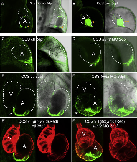
Loss of endocardium and hemodynamic stimulation affects development of the CCS. A: 3 dpf CCS:clo sibling. B: 3 dpf CCS:clom39-/- mutant. SAN CCS cells (arrows) spread widely in clom39-/-. C, E: Control CCS embryos at 2 dpf and 3 dpf, respectively. D, F: tnnt2 morphants at 2 dpf and 3 dpf, respectively. E′, F′: The same embryos as in E and F, showing dsRed expression of CCS:Tg(myl7:dsRed). Dotted white lines outline the chambers of the embryonic zebrafish heart. A-D: lateral view; E-H: ventral view. Abbreviations: A, atrium; SAN, sino-atrial region; V, ventricle. |
Expression Data
| Genes: | |
|---|---|
| Fish: | |
| Knockdown Reagent: | |
| Anatomical Terms: | |
| Stage Range: | Long-pec to Protruding-mouth |
Expression Detail
Antibody Labeling
Phenotype Data
| Fish: | |
|---|---|
| Knockdown Reagent: | |
| Observed In: | |
| Stage Range: | Long-pec to Protruding-mouth |
Phenotype Detail
Acknowledgments
This image is the copyrighted work of the attributed author or publisher, and
ZFIN has permission only to display this image to its users.
Additional permissions should be obtained from the applicable author or publisher of the image.
Reprinted from Gene expression patterns : GEP, 21(2), Poon, K.L., Liebling, M., Kondrychyn, I., Brand, T., Korzh, V., Development of the cardiac conduction system in zebrafish, 89-96, Copyright (2016) with permission from Elsevier. Full text @ Gene Expr. Patterns