
Fig. 3
- ID
- ZDB-FIG-161101-3
- Publication
- Poon et al., 2016 - Development of the cardiac conduction system in zebrafish
- Other Figures
- All Figure Page
- Back to All Figure Page
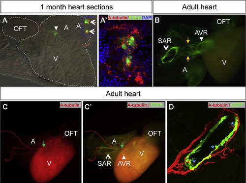
The EGFP expression at the ring of cells at the SAN and AVC is maintained into adulthood in the CCS transgenics. A: A cryosection of a one mpf CCS EGFP + cells stained with anti-acetylated tubulin (red), anti-EGFP (green) and counterstaining with DAPI (blue) shows that the SAR pacemaker cells are neuronally innervated. A′: Enlargement of inset from A - fluorescence at the atrium base. B: In the adult heart, the EGFP expression is localized to the ring of cells located at the SAR and AVR as well as cellular connection between SAN and AVC (labeled by orange arrow). C-C′: Adult whole heart double stained with anti-EGFP (green) and anti-acetylated tubulin (red) shows nerve fibers extending from SAN towards AVC, in close proximity to EGFP cells (green arrows). D: Confocal image of SAN ring showing multiple contacts (blue asterisks) with SAN conduction cells ring. All arrows in this Fig points to SAN EGFP cells whereas arrowheads point to AVC EGFP cells. Dotted white lines in A outline the chambers of the adult zebrafish heart. Abbreviations: A, atrium; AVC, atrio-ventricular region; OFT, outflow tract; SAN, sino-atrial region; SV, sinus venosus; V, ventricle. |
| Gene: | |
|---|---|
| Fish: | |
| Anatomical Terms: | |
| Stage Range: | Days 30-44 to Adult |
Reprinted from Gene expression patterns : GEP, 21(2), Poon, K.L., Liebling, M., Kondrychyn, I., Brand, T., Korzh, V., Development of the cardiac conduction system in zebrafish, 89-96, Copyright (2016) with permission from Elsevier. Full text @ Gene Expr. Patterns