
Fig. S14
- ID
- ZDB-FIG-160916-20
- Publication
- Shibata et al., 2016 - Fgf signalling controls diverse aspects of fin regeneration
- Other Figures
- All Figure Page
- Back to All Figure Page
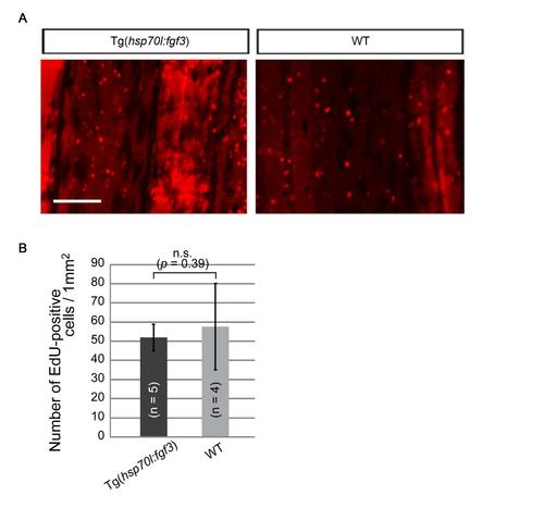
fgf3 overexpression did not affect cell proliferation in uncut fin. (A) EdU incorporation in intact fin from the Tg overexpressing fgf3 and the sibling wild-type (WT) zebrafish. Fish were labelled with EdU for 12 hrs after heat shock. The red colour of fin rays is a non-specific background caused by Edu staining. Scale bar, 200 µm. (B) Quantitative analysis of EdU incorporation in the intact fin. The number of EdU positive cells is divided by the area of fin. The number of EdU-positive cells per unit area is indistinguishable between Tg(hsp70:fgf3) and WT. Error bars represent mean ± s.e.m. Student’s t test was performed to assess statistical significance. n, the number of fish. |