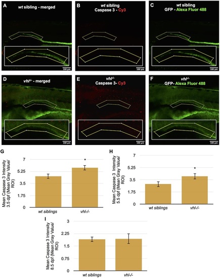
Proximal vhl-/- pronephric tubules have increased cell death at 3.5 and 5.5dpf compared to wt siblings. (A-F) Caspase 3 immunofluorescence was performed in whole-mount 3.5, 5.5 and 8.5dpf Tg(ATPase1.a1A4:GFP) vhl-/- and wt siblings. Caspase 3 staining intensity (red) was then quantified within an ROI (yellow box) encompassing the proximal pronephros (green) using ImageJ. Merged caspase 3 (red) and GFP (green) staining in 5.5dpf wt sibling (A) and vhl-/- (D) larvae. Caspase 3 staining only is shown in 5.5dpf wt sibling (B) and vhl-/- (E). GFP staining only is shown in 5.5dpf wt sibling (C) and vhl-/- (F). (G) Quantification of the mean caspase 3 staining intensity within a ROI in 3.5dpf vhl-/- versus wt siblings, (H) 5.5dpf vhl-/- versus wt siblings and (I) 8.5dpf vhl-/- versus wt siblings. To assist with visualization of caspase 3 staining, the contrast of this image has been adjusted globally using ImageJ software. The quantitative analysis presented was performed on non-enhanced images. Each 3.5, 5.5 and 8.5dpf experiment had ten larvae per sample group and was performed in duplicate. Data represent mean±s.e.m. *P<0.05 (paired two-tailed t-test).
|
