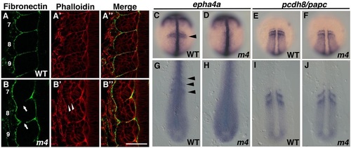
Impaired formation of the morphological somite boundary in the Mesp quadruple mutant. (A,B) Wild-type (A-A′′) and Mesp quadruple mutant (B-B′′) embryos stained with anti-Fibronectin antibody (A,B) and Phalloidin (A′,B′). The numbers shown in A and B indicate the position of each somite from the most anterior one. White arrows indicate gaps of Fibronectin assembly at somite boundaries. White arrowheads indicate disruption of cell arrangement at a somite boundary. (C,D,G,H) Expression of epha4a in wild-type (C,G) and Mesp quadruple mutant (D,H) embryos. Embryos were fixed at the 1-somite stage (C,D) and the 8-somite stage (G,H). Striped expression of epha4a was lost in Mesp quadruple mutant embryos (D: 100%, n=9; H: 100%, n=12). Black arrowheads indicate striped expression of epha4a in the anterior PSM and somites. (E,F,I,J) Expression of papc in wild-type (E,I) and Mesp quadruple mutant (F,J) embryos. Embryos were fixed at the 1-somite stage (E,F) and the 11-somite stage (I,J). Striped expression of papc was not affected by disruption of the function of the four Mesp genes (F: 100%, n=9; J: 100%, n=18). Scale bar: 50µm.
|
