
FIGURE
Fig. 2
- ID
- ZDB-FIG-160908-2
- Publication
- Yabe et al., 2016 - Mesp quadruple zebrafish mutant reveals different roles of mesp genes in somite segmentation between mouse and zebrafish
- Other Figures
- All Figure Page
- Back to All Figure Page
Fig. 2
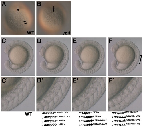
Phenotype of the Mesp quadruple homozygous mutant. (A,B) Dorsal view of wild type and Mesp quadruple homozygous mutant at the 1-somite stage. The arrowhead points to the morphological somite boundary. The arrows indicate the notochord. (C-F) Morphology of wild-type (C), mespaa and mespba double mutant (D), mespab and mespbb double mutant (E), and Mesp quadruple homozygous mutant (m4; F) embryos at the 18-somite stage. Magnified images are also indicated (C′-F′). The bracket indicates the impaired somite boundary in the Mesp quadruple mutant. |
Expression Data
Expression Detail
Antibody Labeling
Phenotype Data
| Fish: | |
|---|---|
| Observed In: | |
| Stage: | 14-19 somites |
Phenotype Detail
Acknowledgments
This image is the copyrighted work of the attributed author or publisher, and
ZFIN has permission only to display this image to its users.
Additional permissions should be obtained from the applicable author or publisher of the image.
Full text @ Development