
Fig. S1
- ID
- ZDB-FIG-160831-13
- Publication
- Xu et al., 2016 - Microglia Colonization of Developing Zebrafish Midbrain Is Promoted by Apoptotic Neuron and Lysophosphatidylcholine
- Other Figures
- All Figure Page
- Back to All Figure Page
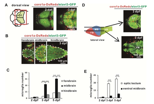
Microglia Distribution in Larval Zebrafish Brain, related to Figure 1 (A) Temporal-spatial distribution of microglia in the brains of 2 dpf and 3 dpf Tg(- 2.8elavl3:eGFP;coro1a:DsRedx) zebrafish embryos. The left panel is the schematic drawing of the dorsal view of zebrafish head region. The optic tectum (OT) is marked in light green. The red square indicates the imaged area of right panels. The right panels show the distribution of coro1a-DsRedx+ microglia (red) inside the elavl3-GFP+ brain (green). (B) Distribution of microglia in the forebrain, midbrain and hindbrain of 3 dpf Tg(- 2.8elavl3:eGFP;coro1a:DsRedx) zebrafish embryos. Red and green signals represent coro1a-DsRedx+ microglia and elavl3-GFP+ neurons respectively. (C) Quantification of microglia number in the forebrain, midbrain and hindbrain of 2 dpf, 3 dpf, and 5 dpf Tg(-2.8elavl3:eGFP;coro1a:DsRedx) embryos. Error bars represent mean SEM. ***: p<0.001. (n=6 for 2 dpf, n=12 for 3 dpf, n=6 for 5 dpf) (D) Distribution of microglia in the optic tectum and ventral midbrain (VM) of 3 dpf Tg(- 2.8elavl3:eGFP;coro1a:DsRedx) zebrafish embryos. The left panel is the schematic drawing of the lateral view of zebrafish brain, which includes the forebrain (FB), midbrain (MB) and hindbrain (HB). The black lines indicate the positions of the coronal optic sections for the optic tectum (upper right panel) and ventral midbrain (VM) (lower right panel). Red and green signals in right panels represent coro1a-DsRedx+ microglia and elavl3-GFP+ neurons respectively. (E) Quantification of microglia number in the optic tectum of the midbrain and the ventral midbrain in Tg(-2.8elavl3:eGFP;coro1a:DsRedx) embryos. Error bars represent mean SEM. ***: p<0.001. (n=6 for 2 dpf, n=12 for 3 dpf, n=6 for 5 dpf). |
Reprinted from Developmental Cell, 38(2), Xu, J., Wang, T., Wu, Y., Jin, W., Wen, Z., Microglia Colonization of Developing Zebrafish Midbrain Is Promoted by Apoptotic Neuron and Lysophosphatidylcholine, 214-22, Copyright (2016) with permission from Elsevier. Full text @ Dev. Cell