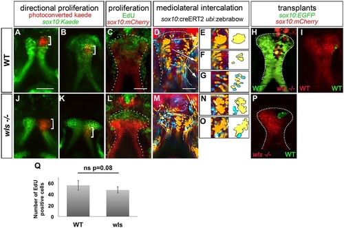
Chondrocytes proliferate normally but fail to intercalate in wls mutants. Anterior is up in all views. (A-I) WT larvae, (J-P) wls mutants. Photoconverted sox10:kaede (red, bracket) in WT (A,B) and wls mutants (J,K) at 55hpf (A,J) and 72hpf (B,K). EdU incorporation assay in WT (C) and wls mutant (L). Multi-spectral clonal analysis in WT (D-G) and wls mutants (M-O). WT chondrocytes undergo a CE from distal to proximal, cells proliferate and group (D,E), converge into columns (D,F) and intercalate (D,G). In wls mutants, cells fail to undergo intercalation (M-O). Yellow and blue groups of cells are illustrated schematically to assist in visualization of the cell intercalation process. (H,I,P) Presumptive cranial NCCs were transplanted from WT sox10:EGFP to wls-/- sox10:mCherry and reciprocally. wls cells intercalated in 4/5 WT (H); WT cells formed groups in 5/6 mutants (P). (I) Control experiment, from WT sox10:EGFP to sox10:mCherry WT. (Q) Quantification of EdU-positive cells. Six WT and five wls mutant embryos were used and positive cells were counted for each. Average of number positive cells were calculated. ns, not significant (Student′s t-test).
|
