
FIGURE
Fig. 2
- ID
- ZDB-FIG-160819-36
- Publication
- Rochard et al., 2016 - Roles of Wnt pathway genes wls, wnt9a, wnt5b, frzb and gpc4 in regulating convergent-extension during zebrafish palate morphogenesis
- Other Figures
- All Figure Page
- Back to All Figure Page
Fig. 2
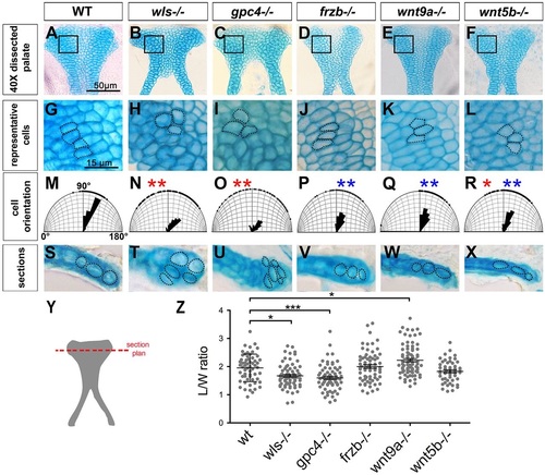
Cell shape and orientation are defective in Wnt signaling mutants. (A-F) Dissected palate (anterior to the top). (G-L) Representative region (as boxed above) magnified to illustrate cell shape and organization. (M-R) Cell orientation was measured and compared with WT (significantly different indicated by red asterisk) and with wls mutant (significantly different indicated by blue asterisk). Watson-U2 test. (S-X) Transverse sections (following the cut plane illustrated in Y) showing chondrocytes stacking in the DV axis. (Z) Graphic representation of the cell L/W ratio. *P<0.05, ***P<0.0001. |
Expression Data
Expression Detail
Antibody Labeling
Phenotype Data
| Fish: | |
|---|---|
| Observed In: | |
| Stage: | Day 4 |
Phenotype Detail
Acknowledgments
This image is the copyrighted work of the attributed author or publisher, and
ZFIN has permission only to display this image to its users.
Additional permissions should be obtained from the applicable author or publisher of the image.
Full text @ Development