
Fig. 7 S1
- ID
- ZDB-FIG-160817-23
- Publication
- Hatzold et al., 2016 - Tumor suppression in basal keratinocytes via dual non-cell-autonomous functions of a Na,K-ATPase beta subunit
- Other Figures
- All Figure Page
- Back to All Figure Page
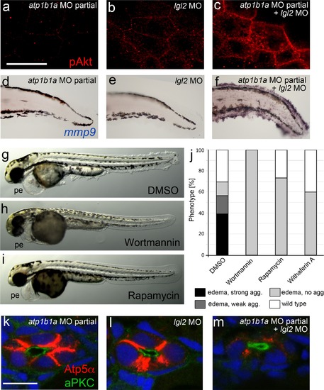
atp1b1a and lgl2 interact genetically to enhance edema formation and AKT phosphorylation and mmp9 expression in basal keratinocytes of embryos raised in hypotonic medium. (a-c) pAKT IF, at 54 hpf, revealing low pAKT levels in embryos injected with low amounts of atp1b1a MO (a) or with lgl2 MO (b), but strongly increased levels in a double-injected embryo (c). For comparison with pAKT levels in wild-type and psoriasis mutants (full loss of Atp1b1a activity), see Figure 8f,g. (d-f). mmp9 WISH, at 54 hpf, revealing low mmp9 expression in embryos injected with low amounts of atp1b1a MO (d) or with lgl2 MO (e), but strongly increased levels in double-injected embryo (f). For comparison with mmp9 expression levels in wild-type and psoriasis mutant, see Figure 9j,j′′. (g-j). Embryos co-injected with sub-phenotypic amounts of atp1b1a MO and lgl2 MO display epidermal aggregates in conjunction with pericardial edema. Treatment with the PI3K inhibitor Wortmannin, the mTORC1 inhibitor Rapamycin or the NFºB inhibitor Withaferin A, starting at 34 hpf, leads to a complete loss of epidermal aggregates, whereas edema persist. (g-i) Representative live images of DMSO-treated controls (g), and of Wortmannin-treated (h) and Rapamycin-treated (i) embryos, at 54 hpf; pe, pericardial edema. (j) Quantification of the phenotypes of control, Wortmannin-, Rapamycin- or Withaferin A-treated embryos as shown in (g- i), at 54 hpf. For classification of phenotypic strengths, see Figure 9a-d. (k-m). IF of Atp5a (red) and aPKC (green), counterstained with DAPI (blue), on transverse sections at 54 hpf, revealing high amounts of Na,K-ATPase α-subunits in the basolateral membrane domains of pronephric duct epithelial cells in embryos injected with low amounts of atp1b1a MO (k) or with lgl2 MO (l), but strongly decreased levels in double-injected embryo (m). Scale bar:10 µm. |