
Fig. S1
- ID
- ZDB-FIG-160714-22
- Publication
- Richardson et al., 2016 - Re-epithelialization of cutaneous wounds in adult zebrafish uses a combination of mechanisms at play during wound closure in embryonic and adult mammals
- Other Figures
- All Figure Page
- Back to All Figure Page
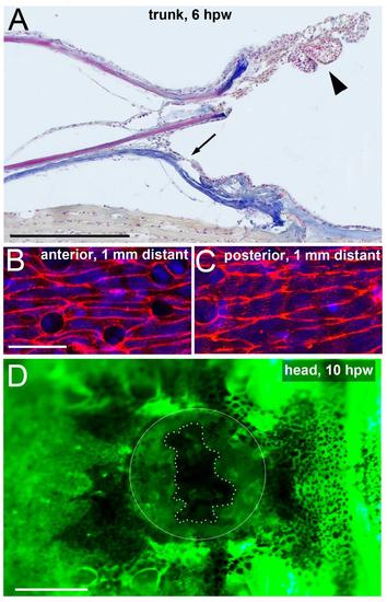
Keratinocytes from both anterior and posterior regions move towards full-thickness wounds. Longitudinal section through a full-thickness wound at 6 hpw, stained with AFOG; only the anterior part of the wound is shown; anterior to the right. Arrow points to anterior border of neo-epidermis formed via re-epithelialization from the posterior side. Arrowhead points to pile of keratinocytes at tip of anterior scale remnant, which have largely lost direct contact to the scale surface (compare with Supplementary Movie 1 and Fig. 1C). (B,C) Phalloidin staining of superficial keratinocytes in regions 1 mm anterior (B) and 1 mm posterior (C) from a full-thickness wound, 4 hpw. Note that on both sides, keratinocytes have elongated in a directed manner (compare with Fig. 4P as negative control). The elongation in anterior regions indicates that these cellular arrangements are largely independent of “pulling” from the LE, as cells pile up at the tip of the scale remnant (see A), rather than re-populating the wound bed. (D) Live fluorescent image of a Tg(krt4:GFP) fish after introduction of a full-thickness wound on the forehead; 10 hpw; anterior to the left; wound encircled; edges of neoepidermis marked by dotted line. In contrast to trunk wounds (compare with Movie 1), the head wound is re-epithelialized from all sides, including the anterior side. Closure of shown wound: 70%; average (n=6): 73±7.7%. Scale bars: A,D: 1 mm; B,C: 20 µm. |