
Fig. 5
- ID
- ZDB-FIG-160714-20
- Publication
- Richardson et al., 2016 - Re-epithelialization of cutaneous wounds in adult zebrafish uses a combination of mechanisms at play during wound closure in embryonic and adult mammals
- Other Figures
- All Figure Page
- Back to All Figure Page
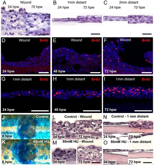
Epidermal cell proliferation regenerates the surrounding epidermis but does not influence re-epithelialization. (A-C) Histological analysis reveals a highly thickened neo-epidermis at both 24 and 72hpw (A). The epidermis 1mm (B) and 2mm (C) distant to the wound is much thinner at 24hpw, but has recovered to normal thickness by 72hpw. (D-I) BrdU-incorporation studies reveal few labelled cells in the wound epidermis from 24hpw to 72hpw (D-F), but a strong increase of BrdU+ cells in the epidermis 1mm distant from the wound (G-I). (J-O) Fish treated with hydroxyurea (HU) exhibit the same rate of re-epithelialization as assessed by Methylene Blue assay at 8hpw (J,K). Histological analysis between control and HU-treated fish reveals similar epidermal thicknesses in the wound at 24hpw and 72hpw (L,M), and 1mm distant from the wound at 24hpw (N,O). However, at 72hpw, the latter has recovered to its normal thickness in the control, but remains thin in the HU-treated fish (N,O). Scale bars: 20µm in A-C, L-O; 50µm in D-J. |