
Fig. 6
- ID
- ZDB-FIG-160706-46
- Publication
- Kawase et al., 2016 - EP300 Protects from Light-Induced Retinopathy in Zebrafish
- Other Figures
- All Figure Page
- Back to All Figure Page
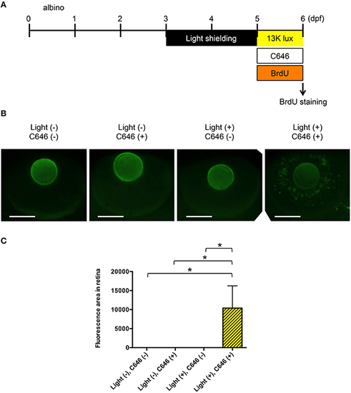
Inhibition of EP300 increases BrdU incorporation in putative Müller cells in the zebrafish model of light-induced retinopathy. (A) Protocol for light-induced retinal damage in larval zebrafish, as described for Figure 4A. After light exposure, whole-mount immunohistochemical staining with anti-BrdU antibody was performed. (B) Representative images of anti-BrdU antibody staining of zebrafish exposed to normal or intense light. Scale bars, 100 µm. (C) Quantitative analysis of BrdU-positive cells in the retinas of zebrafish exposed to the conditions shown in (B). *p < 0.05. Data are the mean ± SEM of n = 8 for all groups. |
| Fish: | |
|---|---|
| Condition: | |
| Observed In: | |
| Stage: | Day 6 |