
|
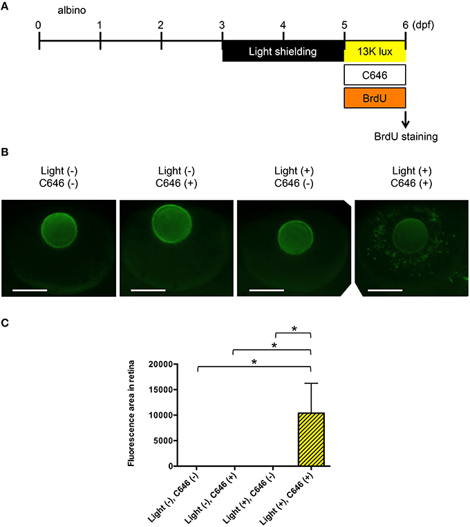
Fig. 6
Inhibition of EP300 increases BrdU incorporation in putative Müller cells in the zebrafish model of light-induced retinopathy. (A) Protocol for light-induced retinal damage in larval zebrafish, as described for Figure 4A. After light exposure, whole-mount immunohistochemical staining with anti-BrdU antibody was performed. (B) Representative images of anti-BrdU antibody staining of zebrafish exposed to normal or intense light. Scale bars, 100 µm. (C) Quantitative analysis of BrdU-positive cells in the retinas of zebrafish exposed to the conditions shown in (B). *p < 0.05. Data are the mean ± SEM of n = 8 for all groups.