
Fig. S3
- ID
- ZDB-FIG-160608-42
- Publication
- Biechl et al., 2016 - Eppur Si Muove: Evidence for an External Granular Layer and Possibly Transit Amplification in the Teleostean Cerebellum
- Other Figures
- All Figure Page
- Back to All Figure Page
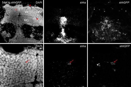
Zebrafish transgenic shh-GFP line brain sections in-situ hybridized for shha at 5 dpf and analyed with confocal microscopy shows shha expression domains as seen in WT (compare to Supplementary Fig. 2) to largely overlap with GFP. Upper row shows overview of valvular cerebellar (Va) level, bottom row is an enlargement. Left panels: DAPI, middle panels: shha, right panels: shhGFP. Red arrowheads indicate border between optic tectum (TeO) and valvula (Va), red circle indicates region of interest. Medulla oblongata is separated from cerebellum by rhombencephalic ventricular space. Importantly, there are dorsal cerebellar cells double-labeled for the transgene (shhGFP) and the in-situ signal (shha, red arrows). |