
Fig. S1
- ID
- ZDB-FIG-160608-40
- Publication
- Biechl et al., 2016 - Eppur Si Muove: Evidence for an External Granular Layer and Possibly Transit Amplification in the Teleostean Cerebellum
- Other Figures
- All Figure Page
- Back to All Figure Page
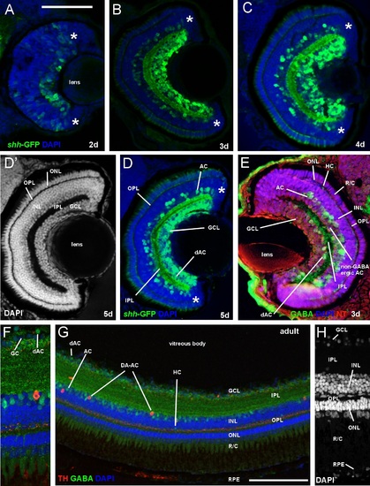
Comparison of retina of zebrafish shh-GFP line with immunostained wildtype larval and adult zebrafish (confocal photomicrographs, except E). (A-D) Sections of retina in transgenic zebrafish at 2, 3, 4, and 5 dpf show GFP-positive amacrine and ganglion cells in addition to fiber net in internal plexiform layer. (D′) Same section as (D) shows retinal layers. Note monolayer of photoreceptors and multiple ganglion cell layers. (E) Wildtype larval zebrafish retina stained immunohistochemically for GABA, visualizing amacrine (incl. some that are displaced into ganglion cell layer) and a few horizontal cells. (FH) Wildtype adult zebrafish retina stained immunohistochemically for GABA and tyrosine hydroxylase (TH) visualizing amacrine cells. Magnification in (F) shows GABAergic amacrine cells in inner nuclear layer and displaced ones (green) next to ganglion cells (blue) and one dopaminergic amacrine (interplexiform) cell (red). Note the latter′s dendritic punctate staining in the inner part of the outer plexiform layer. Magnification in (H) shows cell somata layers with DAPI. Note in inner nuclear layer the lighter stain of amacrine cells compared to bipolar cells and the elongated shape of horizontal cells. Counterstains: DAPI and NeuroTrace (NT). Asterisks: proliferative retinal edge. Scale bar in (A): 100µm (applies to panels B-E), in (G): 50µm. Abbreviations: AC amacrine cells (retina), dAC displaced amacrine cells, DA-AC dopaminergic amacrine (interplexiform) cells, GC ganglion cell, GCL ganglion cell layer, HC horizontal cells, INL inner nuclear layer, IPL inner plexiform layer, ONL outer nuclear layer, OPL outer plexiform layer, R/C rods/cones (inner & outer segments), RPE retinal pigment epithelium |