
FIGURE
Fig. 8
Fig. 8
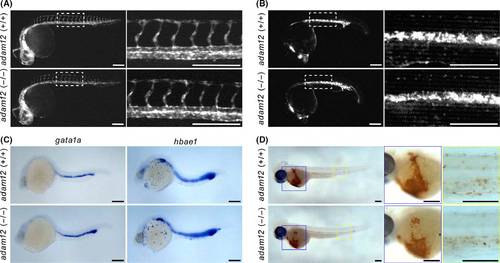
Phenotypic analysis of the blood vessel formation and hematopoiesis. (A) Vascular tube formation and intersegmental vessel (ISV) elongation in fli1a: GFP embryos. DA, dorsal aorta; PCV, posterior cardinal vein at 30-hpf. (B, C) Generation of primitive erythroblasts as visualized by gata1a:mRFP expression (B) and in situ hybridization (C) at 26 hpf. (D) Mature erythrocytes in embryos stained with o-dianisidine at 3-dpf. Scale bar: 200 µm. |
Expression Data
Expression Detail
Antibody Labeling
Phenotype Data
| Fish: | |
|---|---|
| Observed In: | |
| Stage Range: | Prim-5 to Protruding-mouth |
Phenotype Detail
Acknowledgments
This image is the copyrighted work of the attributed author or publisher, and
ZFIN has permission only to display this image to its users.
Additional permissions should be obtained from the applicable author or publisher of the image.
Full text @ Dev. Growth Diff.