
Fig. 1
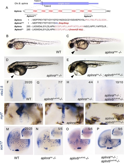
aplnra mutant embryos display defects in endoderm and heart formation. (A) Schematic detailing the aplnramax and aplnrains alleles. TM indicates the transmembrane domain. (B–E) Gross morphology of aplnramax,aplnrains and aplnrains; aplnrbhu4145 mutant embryos compared to WT (wild type) at 48 hpf (hours post-fertilization). (F-I) nkx2.5 expression at the 15 somite stage in WT, aplnrbhu4145, aplnrains, and aplnrains; aplnrbhu4145 mutant embryos. Dorsal view with anterior to the top. (J-L) In situ hybridization showing expression of myl7 at 48 hpf in aplnrains and aplnrains; aplnrbhu4145 embryos compared to WT when viewed from the anterior. (M-P) Comparison of sox17 expression at 8 hpf between WT, aplnrains, aplnrbhu4145and aplnrains; aplnrbhu4145 mutant embryos. Dorsal views are shown with a lateral view in inset panels. |
| Genes: | |
|---|---|
| Fish: | |
| Anatomical Terms: | |
| Stage Range: | 75%-epiboly to Long-pec |
| Fish: | |
|---|---|
| Observed In: | |
| Stage Range: | 75%-epiboly to Long-pec |