
Fig. S4
- ID
- ZDB-FIG-160405-20
- Publication
- Jaffe et al., 2016 - c21orf59/kurly Controls Both Cilia Motility and Polarization
- Other Figures
- All Figure Page
- Back to All Figure Page
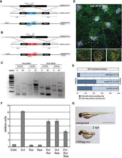
Related to Figure 3. Generation of Xenopus kur Mutants, Tissue-Level Polarity Defects, and Repression of Canonical Wnt Signaling by Kur. (A-C) Schematic diagram of CRISPR/Cas9-induced homologous recombination (HR) knock-in (KI) strategy. A single guide RNA (gRNA) cuts both the long and short kur alleles, then targeting constructs with allele-specific homology arms (HA) are used to integrate BFP (A) and RFP (B), respectively, into the endogenous locus, followed by termination codons and a polyA tail. PCR using a BFP- or RFP-specific primer and a primer outside the region of homology (C) indicated successful KI in F0 mosaic embryos. (D-E) Tissue level polarity is perturbed when kur is depleted. Representative image from a kur MO-treated embryo (ATG MO 2) expressing the rootlet marker GFP-CLAMP (green) and the basal body marker Centrin-RFP (red) stained with anti-acetylated tubulin antibody (purple) (D). Yellow arrows indicate average cilia orientation of individual ciliated cells. Three cells are enlarged without acetylated tubulin staining showing cilia orientation. Quantitation of ciliated cell polarity in control embryos, embryos overexpressing the PCP component vangl2, and kur morphant embryos (E). (F) Luciferase readouts of the Super8XTopFlash reporter. Expression of Dvl alone activates the reporter whereas coexpression of Dvl and Kur or Dvl and Sea reduces activity, which is further reduced by expression of Kur and Sea together. Error bars represent standard deviation. (G) Overexpression of kur by RNA injection at the 1-cell-stage induced moderate dorsalization. |