
FIGURE
Fig. S4
- ID
- ZDB-FIG-160404-15
- Publication
- Koenig et al., 2016 - Vegfa signaling promotes zebrafish intestinal vasculature development through endothelial cell migration from the posterior cardinal vein
- Other Figures
- All Figure Page
- Back to All Figure Page
Fig. S4
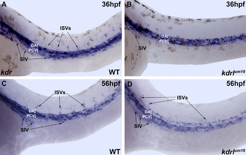
kdr expression is not significantly affected in kdrlum19 embryos. Brightfield microscope images of in situ hybridization staining. kdr is expressed at similar levels in WT (A,C) and kdrlum19 (B,D) embryos at 36 and 56 hpf including the developing SIV (indicated by arrows). |
Expression Data
Expression Detail
Antibody Labeling
Phenotype Data
Phenotype Detail
Acknowledgments
This image is the copyrighted work of the attributed author or publisher, and
ZFIN has permission only to display this image to its users.
Additional permissions should be obtained from the applicable author or publisher of the image.
Reprinted from Developmental Biology, 411(1), Koenig, A.L., Baltrunaite, K., Bower, N.I., Rossi, A., Stainier, D.Y., Hogan, B.M., Sumanas, S., Vegfa signaling promotes zebrafish intestinal vasculature development through endothelial cell migration from the posterior cardinal vein, 115-27, Copyright (2016) with permission from Elsevier. Full text @ Dev. Biol.