
Fig. S1
- ID
- ZDB-FIG-160404-12
- Publication
- Koenig et al., 2016 - Vegfa signaling promotes zebrafish intestinal vasculature development through endothelial cell migration from the posterior cardinal vein
- Other Figures
- All Figure Page
- Back to All Figure Page
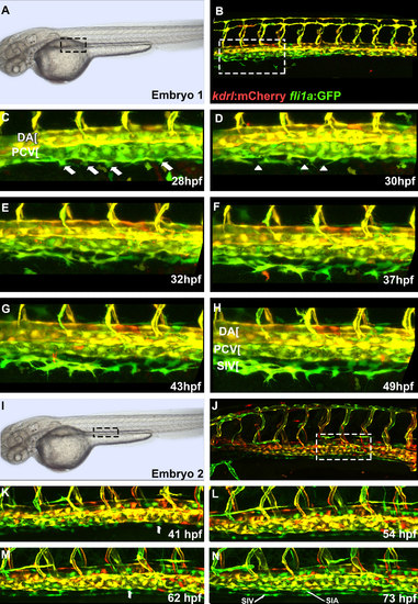
Intestinal vasculature forms through endothelial cell migration from the posterior cardinal vein. Confocal microscope images of live Tg(kdrl:mCherry);Tg(fli1a:EGFP) embryos at 28-48 hpf. (A,B) regions shown in panels C-H are indicated by dashed boxes. (C-H) Endothelial cells begin to migrate out of the posterior cardinal vein at 28 hpf and by 35 hpf begin to coalesce to form the SIV (anterior portion of SIV). (I,J) regions shown in panels K-N are indicated by dashed boxes. (K-N) Cropped image of posterior region of the trunk in another embryo shows cells migrating from the posterior cardinal vein that form both the SIV (K) and SIA (M) as well as connections that form between SIV and SIA. Arrows indicate cells migrating from PCV while arrowheads mark gaps that are closing by extending filopodia. Anterior-left, dorsal-top. |
Reprinted from Developmental Biology, 411(1), Koenig, A.L., Baltrunaite, K., Bower, N.I., Rossi, A., Stainier, D.Y., Hogan, B.M., Sumanas, S., Vegfa signaling promotes zebrafish intestinal vasculature development through endothelial cell migration from the posterior cardinal vein, 115-27, Copyright (2016) with permission from Elsevier. Full text @ Dev. Biol.