
Fig. S1
- ID
- ZDB-FIG-160316-18
- Publication
- Yoshimatsu et al., 2016 - Presynaptic partner selection during retinal circuit reassembly varies with timing of neuronal regeneration in vivo
- Other Figures
- All Figure Page
- Back to All Figure Page
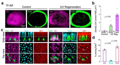
UV cone ablation induces regeneration of specific cone types. (a) Wholemount view of UV cone (magenta) distributions at 15 dpf in a control retina (no ablation) and in a retina after UV cone ablation at 5 dpf Tg(sws1:nfsB-mCherry). EdU (green) was applied to the animals from 1-4 dpa. Dense ring of EdU+ cells mark the ciliary marginal zone where cell genesis persists into adulthood. (b) Densities of EdU+ UV cones at 15 dpf plotted for control retina and retinae with UV cone regeneration (UV Reg.). The density of all UV cones (EdU + or EdU -) present in the UV reg. retina are also plotted. Each circle represents measurements from one retina. (c) (Side view) Orthogonal views of confocal image stacks from control and UV cone ablated retinae showing colabeling for various cone types and EdU (green). zpr1 positive cells (red) are red or green cones. The levels at which UV, blue and red/green cone nuclei stratify are indicated by their respective lines. (Top view) En face view of the cones and EdU labeled nuclei at the levels of the lines marked in the sideviews. Arrowheads point to EdU+ nuclei. (d) Percentage of EdU+ cones that were UV, blue or red/green comprising the regenerated population. |