
Fig. 1
- ID
- ZDB-FIG-160316-13
- Publication
- Yoshimatsu et al., 2016 - Presynaptic partner selection during retinal circuit reassembly varies with timing of neuronal regeneration in vivo
- Other Figures
- All Figure Page
- Back to All Figure Page
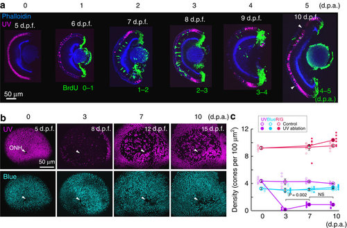
Ultraviolet-cone ablation triggers cone regeneration. (a) Hemisected retina before (at 5 days post fertilization, d.p.f.) and after 1–5 days post ablation (p.d.a.). Ultraviolet cones (magenta) were visualized in Tg(sws1:nfsB-mCherry). BrdU (green) was applied for 1 day at various time points after ablation. The hemisected eyes were stained for phalloidin (blue) to mark in the inner and outer synaptic layers. Arrowheads indicate boundary between original ablated area and the region where ultraviolet cones were added subsequently due to continual retinal growth. (b) Whole-mount retina from Tg(sws1:nfsB-mCherry, sws2:GFP) fish fixed at indicated time points, showing en face view of ultraviolet (magenta) and blue (cyan) cones before and after ultraviolet-cone ablation and regeneration. Arrowheads point to the location of the optic nerve head (ONH). (c) Cone densities before and after ultraviolet-cone ablation, and from age-matched control animals. Large circles are the mean values and small circles indicate values from each retina. Error bars are s.e.m. Red and green cones (R/G) were visualized by immunostaining with the zpr1 antibody. NS, not significant (P=0.94). P values are from Mann–Whitney rank-sum test. UV, ultraviolet. |