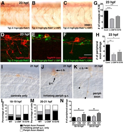
Decreased peripheral axon branching in transgenics expressing CMT2b Rab7 mutants. a-c Lateral views (anterior to the left) of RB neurons labeled with HNK-1 antibody (brown) in 23 hpf transgenic embryos expressing wildtype GFP-Rab7 (a) or GFP-Rab7 L129F (b-c) in all RB neurons, showing decreased branching in Tg(GFP-Rab7L129F) embryos. d-f Mosaic labeling of single RB cells with TagRFP-caax membrane label (red) in 23 hpf transgenic embryos expressing wildtype GFP-Rab7 (d) or GFP-Rab7 L129F (e, f) in all RB neurons. d Red-labeled RB in wildtype GFP-Rab7 transgenic embryo with widely branched peripheral axon. e Red-labeled RB in GFP-Rab7 L129F transgenic embryo does not extend a peripheral axon. c Red-labeled RB in GFP-Rab7 L129F transgenic embryo extends a short peripheral axon that does not branch. Scale bar = 20 µm. g Quantification of peripheral branches crossing horizontal myoseptum in 23 hpf Tg(-3.1ngn:gfp-Rab7) control (Cont) embryos (n = 14 embryos), Tg(-3.1ngn:gfp-Rab7 L129F) embryos (L129F, n = 63 embryos), and Tg(-3.1ngn:gfp-Rab7 K157N) embryos (K157N, n = 20 embryos). ****p < 0.0001, ***p = 0.0001. Unpaired, two-tailed t-test. h Number of peripheral branch tip endings in 23 hpf Tg(-3.1ngn:gfp-Rab7) control (n = 9 cells in 6 embryos), Tg(-3.1ngn:gfp-Rab7 L129F) (n = 24 cells in 24 embryos), or Tg(-3.1ngn:gfp-Rab7 K157N) (n = 21 cells in 15 embryos) embryos is significantly reduced in embryos expressing CMT2b-associated Rab7 mutants. **p = 0.002, *p = 0.04. Unpaired, two-tailed t-test. i-k Lateral views of 21 hpf embryos injected with ngn:TagRFP-caax and labeled with anti-TagRFP antibody. I, Example of RB with central axons only and no peripheral axon. j Example of RB with a peripheral growth cone (g.c.) just initiating (arrow). Cell body (c.b.) is out of focus. k, Example of RB with short peripheral (with 2 endings, arrowheads) extended out of the spinal cord. Cell body (c.b.) is out of focus. l-m Quantification of percentage neurons with peripheral axons at 18–19 hpf (l) and 20–21 hpf (m). There are no significant differences between wildtype and CMT2b Rab7 mutants. At 18–19 hpf: wildtype n = 65 neurons, CMT2b L129F n = 61 neurons, CMT2b K157N n = 10 neurons, p = 0.30 Chi-Square test. At 20–21 hpf: wildtype n = 75 neurons, CMT2b L129F n = 28 neurons, CMT2b K157N n = 27 neurons, p = 0.15 Chi-Square test. n, Analysis of peripheral axon branch endings. At 18–19 hpf and 20–21 hpf, wildtype vs. CMT2b K157N, *p = 0.03 unpaired student’s t-test
|
