
Fig. 3
- ID
- ZDB-FIG-160308-14
- Publication
- Ponomareva et al., 2016 - Charcot-Marie-Tooth 2b associated Rab7 mutations cause axon growth and guidance defects during vertebrate sensory neuron development
- Other Figures
- All Figure Page
- Back to All Figure Page
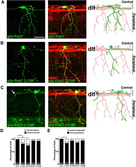
Central axon guidance errors in sensory axons expressing CMT2b Rab7 mutants. a-c Confocal projections (lateral views, anterior to the left) of embryos labeled with HNK-1 antibody (red) and individual RBs expressing GFP-Rab7 forms (green). Drawings at right highlight morphology of one neuron in green. a Wildtype GFP-Rab7 expressing neuron with two central axons traveling in the dorsal longitudinal fascicle (DLF), and with one peripheral axon branching in the skin. b, c RB neurons expressing GFP-Rab7 L129F show lack of outgrowth of ascending central axon (b) or central axon guidance errors (c). Central axon leaving DLF indicated by an arrow in (c). d Quantification of percentage of neurons lacking a central axon. Cont = wildtype Rab7: n = 30 cells in 15 embryos; Rab7 L129F: n = 22 cells in 20 embryos, *p = 0.03; Rab7 K157N: n = 12 cells in 8 embryos, *p = 0.02; Rab7 N161T: n = 16 cells in 11 embryos, p = 0.3; Rab7 V162M: n = 14 cells in 12 embryos, p = 0.3; Fisher’s exact tests. e Quantification of percentage of cells with central axon guidance errors. Cont = wildtype Rab7: n = 30 cells in 15 embryos; Rab7 L129F: n = 24 cells in 19 embryos, *p = 0.03; Rab7 K157N: n = 13 cells in 9 embryos, p = 0.3; Rab7 N161T: n = 13 cells in 11 embryos, p = 0.09; Rab7 V162M: n = 14 cells in 12 embryos, p = 0.3. Scale bar = 40 µm |