
Fig. 3
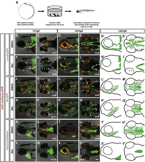
tcf21+ cells Are Required for Head Muscle Formation but Not for Head Vasculature Formation (A) Schematic representation of tcf21+ cell ablation experiment. Images modified from [29]. (B–Y′) Live images and schematic diagrams of embryos of indicated stages carrying indicated transgenes treated with the vehicle control DMSO or Mtz as depicted in (A). Note that in (F)–(I), the prechordal plate-derived extraocular muscles (arrows in F–I) and the somite-derived sternohyoideus muscles (asterisks in F–I, H′, and I′) are not ablated. Arrows in (J–Q, L′, and M′) indicate the HA; arrowheads point to the loop formed by the PAA 1 and the opercular artery, and numbers indicate PAAs 3–6. Note that the HA is missing in the tcf21+ cell-ablated embryos. Note that in (V)–(Y) (schematic diagrams X′ and Y′), all nkx2.5+ cells outside of the heart and pericardium are ablated. The light green color in (T2) and (U2) marks the nkx2.5+ head muscles. The nkx2.5+ endothelial cells are indicated by dark green. The scale bar represents 100 µm. Anterior is to the left. Lateral views (first, third, and fifth column) and ventral views (second, fourth, and sixth column). Sample number of imaged embryos is indicated as a total for the ventral and lateral views. See also Figure S1. |
| Genes: | |
|---|---|
| Fish: | |
| Condition: | |
| Anatomical Terms: | |
| Stage Range: | Protruding-mouth to Day 5 |
| Fish: | |
|---|---|
| Condition: | |
| Observed In: | |
| Stage Range: | Protruding-mouth to Day 5 |