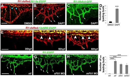
Notch activity is required for final remodeling of the subintestinal plexus but is dispensable for its initial development. (A-D) The subintestinal plexus of 72hpf Tg(fli1:dsRed;flt1_9a_cFos:GFP) embryos treated with DAPT between 24 and 72hpf shows the presence of ectopic leading buds (B,C, arrows; D), malformed SIA (B, asterisks) and active filopodia along the SIV (C, arrows). nDMSO=29; nDAPT=36. ***P<0.001. (E) Spatiotemporal characterization of Notch signaling activation during development of the subintestinal plexus, as detected in Tg(fli1:dsRed;12xNRE:EGFP) double transgenic embryos. EGFP is detected in the SIA starting at 50hpf (arrowheads) and in the leading buds of 60hpf embryos (arrow). (F-I) Downregulation of flt1 results in ectopic leading buds along the SIV of stl mutants (F, arrows), sFlt1 (G, arrows) and mFlt1 (H, arrows) morphants, quantified in I. Yellow channel denotes colocalization of green and red fluorescence. Scale bars: 50µm. nWT=16, nstl=13, nmFlt=27, nsFlt=18. ***P<0.001. Error bars represent s.e.m.
|
