
Fig. 2
- ID
- ZDB-FIG-160205-55
- Publication
- Nikolaou et al., 2015 - Lamination Speeds the Functional Development of Visual Circuits
- Other Figures
- All Figure Page
- Back to All Figure Page
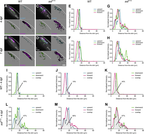
Lamination of DS-RGC Axons in the Tectum Is Lost in astray Mutants (A–D) Composite parametric maps generated from all Tg(Isl2b:Gal4;UAS:SyGCaMP3) fish imaged showing the spatial distribution of DS-RGC subtypes in the tectal neuropil of WT larvae (n = 8, total of 24 optical sections) at 4 dpf (A) and 7 dpf (B), and astti272z larvae (n = 9, total of 27 optical sections) at 4 dpf (C) and 7 dpf (D). Voxel brightness is proportional to the summed incidence of each functional subtype across all larvae imaged. The standard space template image derived from the mean fluorescence image of SyGCaMP3-expressing axons (grayscale) provides an anatomical reference. Dashed lines indicate the position of the skin overlaying the tectum. Scale bar represents 20 µm. A, anterior; L, lateral; SFGS, stratum fibrosum et griseum superficiale. (E–H) Line plots generated from the composite parametric maps in (A)–(D) illustrating the distribution of DS-RGC subtypes along the laminar axis of the tectum in WT larvae at 4 dpf (E) and 7 dpf (F), and astti272z larvae at 4 dpf (G) and 7 dpf (H). Line plots show normalized intensity of each DS-RGC subtype as a function of its distance from the skin. (I–N) Pairwise comparisons showing the degree of spatial overlap between upward and downward (I and L), upward and forward (J and M), and downward and forward (K and N) DS-RGC subtypes within WT (I–K) and astti272z (L–N) tecta at 4 dpf. Dotted area represents the area of intersection between the two subtypes. Values shown represent the fraction of downward (for I and L) or forward (for J, K, M, and N) DS voxels that spatially overlap with upward or downward DS voxels. See also Figures S2 and S3. |