
Fig. 1
- ID
- ZDB-FIG-160205-25
- Publication
- Chen et al., 2015 - A Zebrafish Model for Studies on Esophageal Epithelial Biology
- Other Figures
- All Figure Page
- Back to All Figure Page
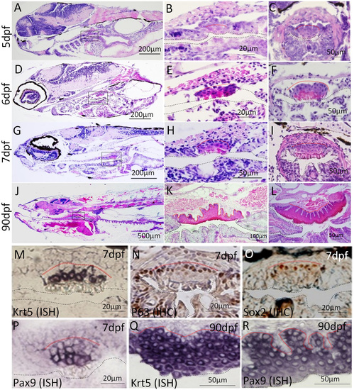
Identification of a non-keratinized stratified squamous epithelium in zebrafish upper digestive tract.(A-L) H&E staining of paraffin sections of zebrafish at 5, 6, 7 and 90 dpf shows the histogenesis of the squamous epithelium. A, D, G and J are the sagittal sections of the whole fish. B, E, H and K are magnifications of the areas in the yellow rectangles in A, D, G and J. Transverse sections show the histology of the squamous epithelium at 5, 6, 7 and 90 dpf (C, F, I, L). ISH for Krt5 (M, Q), IHC for P63 (N), IHC for Sox2 (O) and ISH for Pax9 (P, R) on transverse sections at 7dpf show the expression of esophageal genes in developing and adult zebrafish. All the pictures are dorsal side up. Base membrane of the squamous epithelium is marked with red dotted line and esophageal lumen is lined with black dotted line. |
| Genes: | |
|---|---|
| Antibody: | |
| Fish: | |
| Anatomical Term: | |
| Stage Range: | Days 7-13 to Adult |