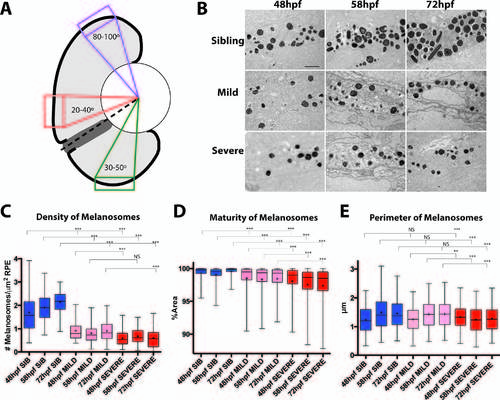
Melanosome formation and maturation is disrupted in nsfbau18 mutants. (A) Cartoon schematic showing the dorsal, central, and ventral regions of the RPE that were analyzed. RPE between 20° and 40° clockwise from an imaginary line (dotted line) drawn between the center of the lens and the center of the optic nerve was defined as central, whereas RPE 80° to 100° was defined as dorsal. Finally, RPE contained within a box 30° to 50° counterclockwise was defined as ventral. (B) Representative TEM images of central RPE for sibling, mild, and severe nsfbau18 RPE at 48, 58, and 72 hours after fertilization. Images oriented distal (choroid) up, proximal (retina) down. Scale bar: 1 µm. (C) Quantification of the average number of melanosomes per square micrometer of RPE indicates that nsfbau18 mutants contained significantly fewer melanosomes than sibling at all time points, and severe nsfbau18 mutants contained significantly fewer melanosomes than mild nsfbau18 mutants at 48 and 72 hpf. (D) The graph depicting the average percentage of melanosomes containing pigment reveals that nsfbau18 melanosomes were significantly less mature than sibling at all time points and that severe mutants were less mature than mild. (E) Graph depicting the average perimeter of melanosomes. Although the average perimeter of melanosomes in sibling and mild nsfbau18 mutants was not significantly different, severe nsfbau18 melanosomes were significantly smaller than sibling. Furthermore, although melanosomal perimeter increased in sibling and mild RPE, severely affected mutant melanosomes decreased over time. (Mann-Whitney U test, *P < 0.01, **P < 0.001, ***P < 0.0001).
|
