|
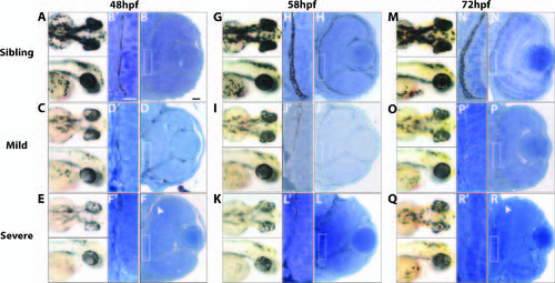
The RPE is disrupted in au18 mutants. Images of sibling and mutant embryos show that the RPE of au18 embryos is hypopigmented at 48 (A, C, E), 58 (G, I, K), and 72 hpf (M, O, Q). Transverse histologic sections of sibling and mutant embryos at 48 (B, D, F) 58 (H, J, L), and 78 hpf (N, P, R) reveal that au18 eye is microphthalmic, but overall retina structure appears to be normal. Areas devoid of pigment are marked by white arrowheads. Scale bar: 20 µm. (B2, D2, F2, H2, J2, L2, N2, P2, R2) Magnified images of RPE show that RPE proximal to the optic nerve was consistently depigmented in au18 and confirm that au18 RPE appears to have fewer melanosomes than wild-type siblings. The posterior lens also appears to be cataractous in severe au18 mutants (F, L, R). Scale bar: 10 µm.
|