
Fig. 5
- ID
- ZDB-FIG-151229-18
- Publication
- Ariotti et al., 2015 - Modular Detection of GFP-Labeled Proteins for Rapid Screening by Electron Microscopy in Cells and Organisms
- Other Figures
- All Figure Page
- Back to All Figure Page
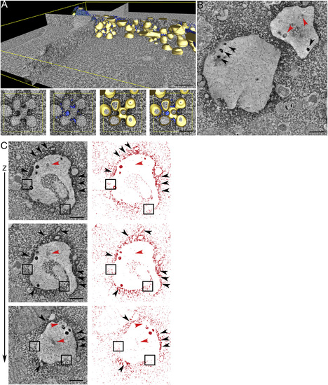
Rapid Screening of a GFP-Tagged Phosphoinositide Probe Library using APEX-GBP Allows for Fast and Reliable Protein Localization (A–H) GFP-PLC-PH, GFP-BTK-PH, GFP-AKT-PH, GFP-TAPP1-PH, GFP-2xFYVEhrs, GFP-OSBP-PH, GFP-2ML1N and GFP-2ML1N (7Q) detected at known subcellular localizations by APEX-GBP co-transfection. Scale bars represent 1 µm. Untransfected adjacent cells without reaction product. Arrowheads represent enriched electron density at the PM, arrows highlight accumulation of reaction product within endosomes. Cy, cytoplasm; PM, plasma membrane; EEs, early endosomes; LE, late endosomes; Golgi, Golgi complex; M, mitochondria; CCP, clathrin-coated pit. The above localizations were confirmed by confocal microscopy; see Figure S3. Comparisons of ultrastructure between modular and directly APEX-tagged PI probes were performed; see Figures S4A–S4C. |
Reprinted from Developmental Cell, 35(4), Ariotti, N., Hall, T.E., Rae, J., Ferguson, C., McMahon, K.A., Martel, N., Webb, R.E., Webb, R.I., Teasdale, R.D., Parton, R.G., Modular Detection of GFP-Labeled Proteins for Rapid Screening by Electron Microscopy in Cells and Organisms, 513-25, Copyright (2015) with permission from Elsevier. Full text @ Dev. Cell