
FIGURE
Fig. 2
- ID
- ZDB-FIG-151229-15
- Publication
- Ariotti et al., 2015 - Modular Detection of GFP-Labeled Proteins for Rapid Screening by Electron Microscopy in Cells and Organisms
- Other Figures
- All Figure Page
- Back to All Figure Page
Fig. 2
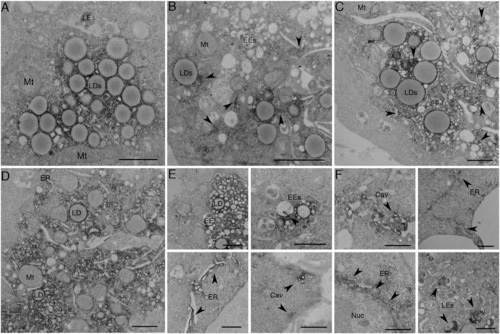
APEX-GBP Allows for Time-Resolved Protein Redistribution (A–F) BHK cells were transfected and serum starved for (A) 0 min, (B) 30 min, (C) 1 hr, (D) 2 hr, (E) 4 hr, and (F) 8 hr to track the redistribution of GFP-CavDGV by APEX-GBP in a time-resolved manner. Arrows highlight regions where electron density has re-distributed from lipid droplets to other intracellular organelles. LD, lipid droplet; Mt, mitochondria; EE, early endosome; LE, late endosome; ER, endoplasmic reticulum; Nuc, Nucleus; Cav, Caveolae. Scale bars represent 500 nm. The above localizations were confirmed by confocal microscopy; see Figure S2. |
Expression Data
Expression Detail
Antibody Labeling
Phenotype Data
Phenotype Detail
Acknowledgments
This image is the copyrighted work of the attributed author or publisher, and
ZFIN has permission only to display this image to its users.
Additional permissions should be obtained from the applicable author or publisher of the image.
Reprinted from Developmental Cell, 35(4), Ariotti, N., Hall, T.E., Rae, J., Ferguson, C., McMahon, K.A., Martel, N., Webb, R.E., Webb, R.I., Teasdale, R.D., Parton, R.G., Modular Detection of GFP-Labeled Proteins for Rapid Screening by Electron Microscopy in Cells and Organisms, 513-25, Copyright (2015) with permission from Elsevier. Full text @ Dev. Cell