
Fig. S2
- ID
- ZDB-FIG-151208-32
- Publication
- Tsujimura et al., 2015 - Spatially differentiated expression of quadruplicated green-sensitive RH2 opsin genes in zebrafish is determined by proximal regulatory regions and gene order to the locus control region
- Other Figures
- All Figure Page
- Back to All Figure Page
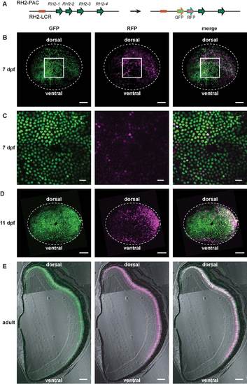
Simultaneous recapitulation of the RH2-1 and RH2-2 expression by the GFP and RFP reporters. (A) GFP and RFP reporters were inserted in the place of RH2-1 and RH2-2, respectively, in the RH2-PAC clone. (B-E) The reporter expression was analyzed in the transgenic fish carrying the RH2-PAC construct of (A). The GFP signals appear as green and the RFP signals appears as magenta. The overlay of the two signals appears as white. The right panels are the overlays of the left and the middle panels. (B) A whole mount retina of a 7-dpf fish. Images are superimposed view of stacked sectional images serially obtained using confocal laser scanning microscopy. The dorsal side is at the top. (C) Expanded views of the central area of a whole mount retina shown in (B). (D) A whole mount retina of an 11-dpf fish as shown in (B). (E) A transverse section of the adult retina. The dorsal side is at the top and the ventral side is at the bottom. Scale bars = 50 µm (B, D), 10 µm (C) and 100 µm (E). |