Fig. 2
- ID
- ZDB-FIG-151208-29
- Publication
- Tsujimura et al., 2015 - Spatially differentiated expression of quadruplicated green-sensitive RH2 opsin genes in zebrafish is determined by proximal regulatory regions and gene order to the locus control region
- Other Figures
- All Figure Page
- Back to All Figure Page
|
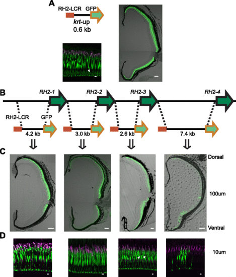
GFP expression patterns specified by the RH2 upstream sequences. a The promoter of keratin 8 attached with the RH2-LCR was linked to a GFP reporter (top left). The GFP was mostly expressed in the SDCs with some ectopic expression as indicated by a white arrowhead (bottom left). The transverse sections of the retinas of the adult transgenic zebrafish showed GFP expression in the entire region from the dorsal to the ventral retina (right). Scale bars = 10 µm (bottom left), 100 µm (right). b Schematic representation of the RH2 upstream constructs with the RH2-LCR. c Images of the transverse sections of the retinas of the adult transgenic zebrafish possessing the respective constructs. The dorsal side is at the top and the ventral side is at the bottom. d Vertical sections of the photoreceptor layer in the same adult retinas as in (c). Immunostaining signals of SDCs by the anti-RH2 antibody appear as magenta, GFP fluorescent signals appear as green, and overlap of the two signals appears as white. Note the weak ectopic expression by the keratin 8 and RH2-3 upstream construct in non-SDC photoreceptor cells as indicated by white arrowheads (a, d). Scale bars = 100 µm (c) and 10 µm (d) |

