
Fig. 4
- ID
- ZDB-FIG-151119-32
- Publication
- Walton et al., 2015 - The Macrophage-Specific Promoter mfap4 Allows Live, Long-Term Analysis of Macrophage Behavior during Mycobacterial Infection in Zebrafish
- Other Figures
- All Figure Page
- Back to All Figure Page
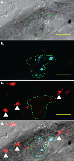
mpeg1-mediated fluorophore expression is attenuated in infected cells. Confocal image of a larval granuloma, 5 days post-infection. The region containing infected cells is indicated by the green dashed line. Within each cell, M. marinum expressing the Cerulean fluorescent protein (cyan signal) are clearly visible. In addition, faint tdTomato fluorescence is visible within the infected cells. White arrowheads indicate examples of bright, uninfected cells exhibiting normal levels of tdTomato fluorescence. a. Brightfield detailing the granuloma in which mpeg1 fluorescent protein expression is attenuated. b. Fluorescent M. marinum in the same area c. Macrophages expressing the tdTomato fluorescent protein. d. Merged image of a-c. Scale bars = 50 µm. |