
FIGURE
Fig. S2
- ID
- ZDB-FIG-151113-7
- Publication
- Xu et al., 2015 - Temporal-Spatial Resolution Fate Mapping Reveals Distinct Origins for Embryonic and Adult Microglia in Zebrafish
- Other Figures
- All Figure Page
- Back to All Figure Page
Fig. S2
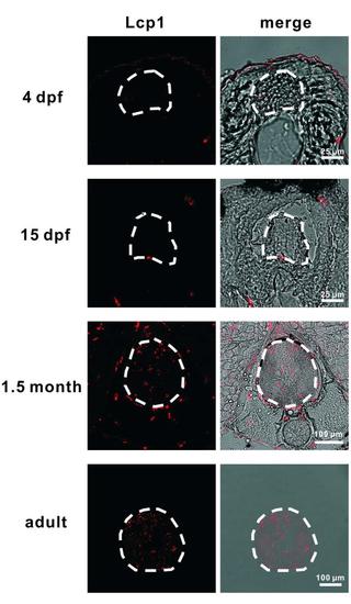
Microglia Colonize the Spinal Cord during Zebrafish Development. Lcp1+ microglia are detected in the spinal cord transverse sections of 1.5 month and adult fish but not of the 4 dpf and 15 dpf fish. Left panels are Lcp1+ signal and right panels represent merged views of Lcp1+ signal and bright field. The spinal cord is outlined by dashed lines. |
Expression Data
Expression Detail
Antibody Labeling
Phenotype Data
Phenotype Detail
Acknowledgments
This image is the copyrighted work of the attributed author or publisher, and
ZFIN has permission only to display this image to its users.
Additional permissions should be obtained from the applicable author or publisher of the image.
Reprinted from Developmental Cell, 34, Xu, J., Zhu, L., He, S., Wu, Y., Jin, W., Yu, T., Qu, J.Y., Wen, Z., Temporal-Spatial Resolution Fate Mapping Reveals Distinct Origins for Embryonic and Adult Microglia in Zebrafish, 632-641, Copyright (2015) with permission from Elsevier. Full text @ Dev. Cell