
Fig. S5
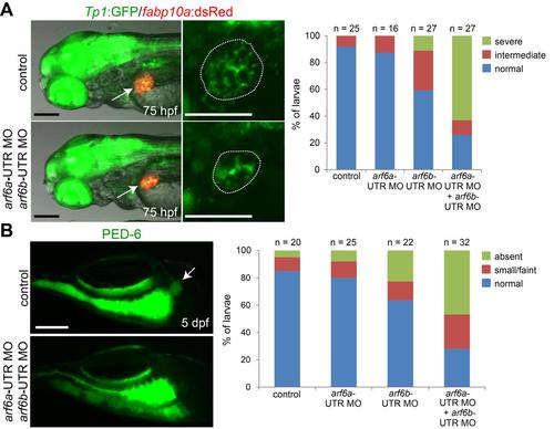
Simultaneous knockdown of arf6a and arf6b results in more severe biliary defects than their single knockdown. (A) The Tg(Tp1:GFP), Tg(fabp10a:dsRed), and Tg(ins:dsRed) lines were used to reveal the intrahepatic biliary structure, the liver, and the dorsal pancreas, respectively. Epifluorescence images showing the expression of these transgenes revealed that a severe biliary defect was observed more often in larvae co-injected with 3 ng of arf6a-UTR and 3 ng of arf6b-UTR MOs than in singly injected larvae. Based on the severity of the biliary defect, larvae were divided into three groups: normal, intermediate, and severe. Graph showing the percentage of larvae in each group. Arrows point to the liver; arrowheads point to the dorsal pancreas. Dotted lines outline the liver. Lateral views, anterior to the left. (B) Epifluorescence images showing PED-6 accumulation in the gallbladder. Based on PED-6 levels in the gallbladder, larvae were divided into three groups: absent, small/faint, and normal. Graph showing the percentage of larvae in each group. Arrows point to the gallbladder. Lateral views, anterior to the right. n indicates the number of larvae examined. Scale bars, 100 µm. |
| Genes: | |
|---|---|
| Fish: | |
| Knockdown Reagents: | |
| Anatomical Terms: | |
| Stage: | Protruding-mouth |
| Fish: | |
|---|---|
| Knockdown Reagents: | |
| Observed In: | |
| Stage Range: | Protruding-mouth to Day 5 |