
Fig. 3
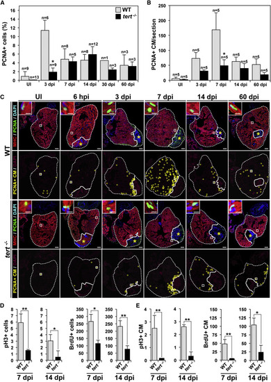
The Absence of Telomerase Severely Affects Cardiomyocyte Proliferation (A and B) Cardiac cells positive for PCNA in UI WT and tert-/- zebrafish hearts and in hearts at the indicated days after cryoinjury. (A) PCNA+ total cardiac cells. (B) PCNA+ cardiomyocytes. Data are means ± SEM of the percentage of PCNA+ cells (A) or number of PCNA+ cardiomyocytes per cardiac ventricle section (at least 3 sections/animal from the indicated number of animals) (B). p < 0.05 compared with WT samples (Mann-Whitney test). (C) WT and tert-/- heart sections immunostained with anti-MHC to mark cardiomyocytes (red) and anti-PCNA to mark cells in S phase (green). Nuclei were counterstained with DAPI (blue). The bottom rows show the location of PCNA+ cardiomyocytes during regeneration (yellow circles). The nuclear area is shown in magenta. Dotted lines outline the ventricle and injured area. Asterisks mark the initial injury site. Insets show high-magnification views of representative PCNA+ cardiomyocytes in the boxed areas. Scale bars, 100 µm (whole-heart views) and 10 µm (magnifications). (D and E) Quantification of pH3+ and BrdU-labeled cardiac cells (D) and cardiomyocytes (E) at 7 and 14 dpi. Data are means ± SEM. p < 0.05, p < 0.01 (unpaired Student’s t test). See also Figures S4 and S5. |