
Fig. 3
- ID
- ZDB-FIG-151008-8
- Publication
- Sallin et al., 2015 - γ-tubulin is differentially expressed in mitotic and non-mitotic cardiomyocytes in the regenerating zebrafish heart
- Other Figures
- All Figure Page
- Back to All Figure Page
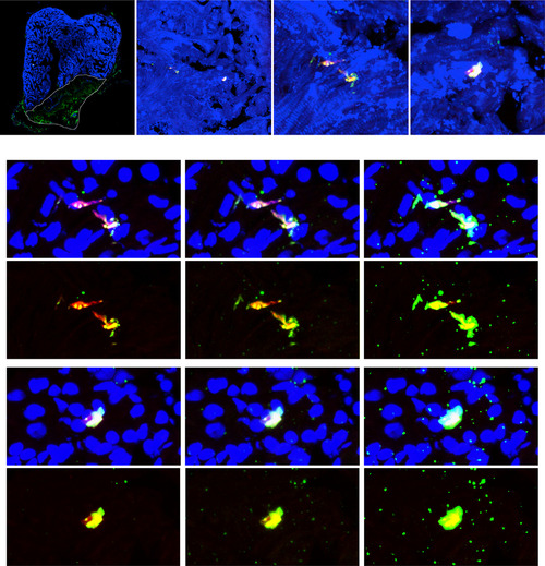
Additional examples of differential γ-tubulin expression in mitotic and non-mitotic cells. (A) Heart section of cmlc2::EGFP transgenic zebrafish at 14 days post cryoinjury (dpci) labeled with antibodies against GFP (cmlc2::EGFP, anti-GFP, cardiac cells, blue), phospho-(Ser10)-histone H3 (PH3, mitosis, red) and γ-tubulin (centrosomes, spindle apparatus, green). Cryoinjured part is encircled with a dashed line. (B) Higher magnification of the framed area shown in (A). (C, D) Higher magnification of the framed areas shown in (B) showing different PH3-positive cells. (C′, D′) The same areas as in (C) and (D) but contrastained with DAPI (blue). The differential subcellular expression of γ-tubulin in mitotic and non-mitotic cells was observed in several regions of each heart. N=5 hearts; Scale bar (A, B, C, C′)=50 µm; S=shadows; M= midtones; H=highlights. |
| Gene: | |
|---|---|
| Antibody: | |
| Fish: | |
| Condition: | |
| Anatomical Terms: | |
| Stage: | Adult |