|
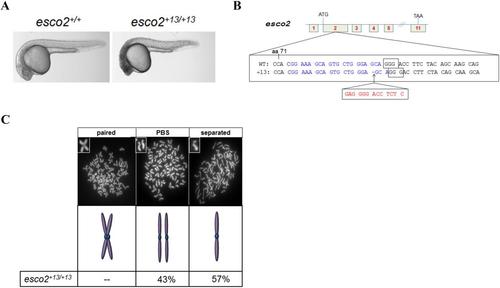
CRISPR/Cas9 derived esco2 null allele recapitulates esco2 retroviral insertion mutant. (A) Brightfield images of 24 hpf esco2+13/+13 allele recapitulates embryonic lethality and head necrosis (n=41 with head necrosis and 137 WT morphology) of esco2m/m. (B) Allele diagram of esco2 +13 allele. A 13 bp insertion occurs in the second ATG containing exon of the 11 exon gene. ATG denotes the transcriptional start site and TAA denotes translation stop site. Blue text indicates CRISPR target site, the PAM is boxed, and red text indicates 13 bp insertion. Codons separated by spaces, note alternative codons in the +13 allele due to the frame shift. (C) Metaphase chromosome spreads from 24 hpf pooled (n=30) esco2+13/+13 embryos. Percentage distribution of each category: “paired”, “paired but separated” (PBS), and “separated”. Representative spreads and diagrams to describe category shown above percentages (n=30 spreads).
|