
FIGURE
Fig. S2
Fig. S2
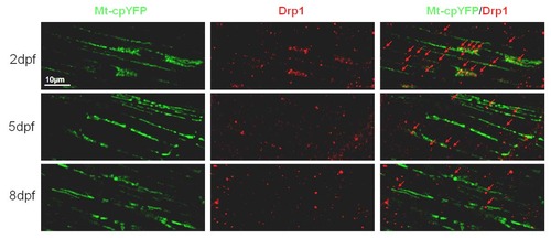
Drp1 were decreasing during zebrafish skeletal muscle development. Note that Drp1 was recruited overlapped with or between Tg(β-actin:mt-cpYFP)-positive mitochondria, decreasing from 2-dpf (n=18), 5-dpf (n=15) to 8-dpf embryos (n=14). Due to severe cpYFP signal loss after fixation, we only investigated mt-cpYFP signals in white fibers. |
Expression Data
| Gene: | |
|---|---|
| Antibody: | |
| Fish: | |
| Anatomical Terms: | |
| Stage Range: | Long-pec to Days 7-13 |
Expression Detail
Antibody Labeling
Phenotype Data
Phenotype Detail
Acknowledgments
This image is the copyrighted work of the attributed author or publisher, and
ZFIN has permission only to display this image to its users.
Additional permissions should be obtained from the applicable author or publisher of the image.
Full text @ PLoS One