
FIGURE
Fig. S5
- ID
- ZDB-FIG-150921-36
- Publication
- Anderson et al., 2015 - In vivo Modeling Implicates APOL1 in Nephropathy: Evidence for Dominant Negative Effects and Epistasis under Anemic Stress
- Other Figures
- All Figure Page
- Back to All Figure Page
Fig. S5
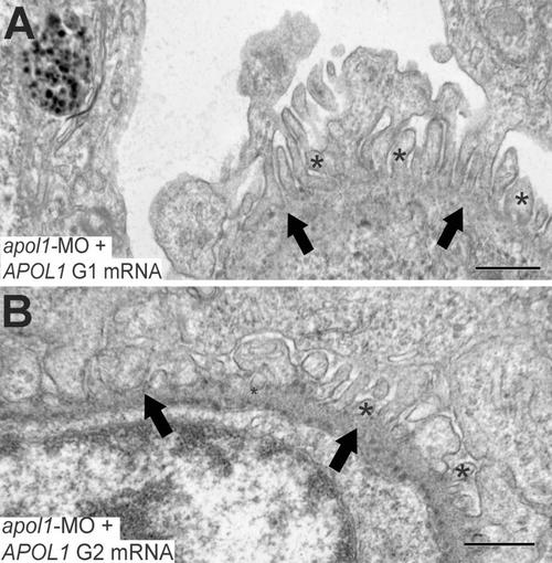
Glomerular ultrastructure of apol1 morphants complemented with human risk alleles. Transmission electron microscopy of zebrafish larval glomeruli imaged at 5 dpf. (A, B) apol1 morphants complemented with risk alleles, G1 and G2 do not rescue the observed defects caused by apol1 suppression, with naked patches of glomerular basement membrane and microvillus processes apparent. *, microvillus protrusions; filled arrowheads, glomerular basement membrane. Scale bars, 500nm. |
Expression Data
Expression Detail
Antibody Labeling
Phenotype Data
Phenotype Detail
Acknowledgments
This image is the copyrighted work of the attributed author or publisher, and
ZFIN has permission only to display this image to its users.
Additional permissions should be obtained from the applicable author or publisher of the image.
Full text @ PLoS Genet.