
Fig. S5
- ID
- ZDB-FIG-150915-65
- Publication
- Gao et al., 2015 - TopBP1 Governs Hematopoietic Stem/Progenitor Cells Survival in Zebrafish Definitive Hematopoiesis
- Other Figures
- All Figure Page
- Back to All Figure Page
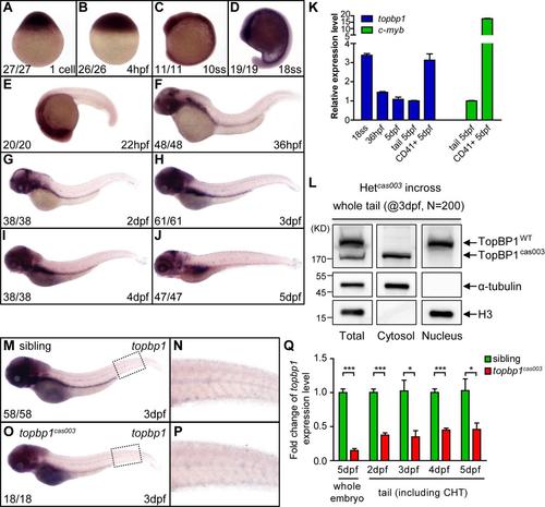
The topbp1 gene is ubiquitously expressed in the development. (A-J) WISH results of topbp1 from 1-cell stage to 5dpf showing global expression of topbp1. ss, somites. The penetrance of the indicated phenotype is shown in the bottom left of each panel. (K) Quantitation of topbp1 in the whole embryos, tails and sorted CD41+ cells at the indicated stage. topbp1 is 3-fold enriched in CD41+ cells within the tail region of Tg(CD41: EGFP) line at 5dpf, demonstrating the expression of topbp1 in HSPCs. c-myb is used as a positive control. (L) Western blotting analysis on endogenous TopBP1WT/TopBP1cas003 protein in cytoplasmic and nuclear fractions of pooled 3dpf embryos from heterozygotes incrossing. TopBP1WT localized in nucleus, but TopBP1cas003 localized in cytosol. (M-P) WISH analysis of topbp1 in sibling and topbp1cas003 mutant embryos at 3dpf. The expression of topbp1 is decreased in mutant, especially in cranial region. (N, P) Enlarged detail of c-myb WISH analysis in CHT region. (Q) Quantitative PCR analysis on the topbp1 mRNA level in the whole embryos at 5dpf or the tails including CHT from 2dpf to 5dpf. The expression level of topbp1 is decreased in the topbp1cas003 mutants. Error bars represent SEM; * represents p<0.05; *** represents p<0.001. |
| Gene: | |
|---|---|
| Fish: | |
| Anatomical Terms: | |
| Stage Range: | 1-cell to Day 5 |