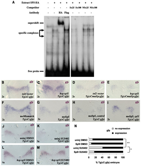
Sp5 factors mediate Fgf signalling on enhancer C. (A) Gel retardation analysis of the binding of an HA-tagged SP5 protein to an oligonucleotide carrying the Sp site from element C. Antibodies against HA or Flag, and specific (S) or non-specific (NS) oligonucleotide competitors (25- or 100-fold molar excess with respect to the probe) were added as indicated. The positions of specific complexes (arrows), a supershifted complex and the free probe are indicated. (B-E) Tg(cC:gfp) or Tg(cCmutSp:gfp) embryos were injected either with a control plasmid (tol2 vector) or an sp5l expression vector (hsp:sp5l) and heat-shocked for 10min at 95% epiboly. ISH for gfp was performed at 2s for B-I. (F,G) Tg(cC:gfp) embryos were injected with morpholinos against sp5l and bts1 (G), or with morpholinos against these genes, but carrying mismatches (F) (four independent experiments). (H,I) Tg(cC:gfp) embryos were co-injected with morpholinos against sp5l and bts1, and with Cherry (H) or Sp5l (I) mRNAs. (J-N) Tg(cC:gfp) embryos were treated with DMSO (J,L) or SU5402 (K,M) from the shield stage onwards and subjected to gfp ISH at 1s. (N) Distribution of Tg(cC:gfp) embryos according to the expression of gfp. The significance of differences was evaluated by Fisher′s Exact Test: *P<0.05; **P<0.02.
|
