
FIGURE
Fig. S4
- ID
- ZDB-FIG-150804-7
- Publication
- Panza et al., 2015 - Live imaging of endogenous protein dynamics in zebrafish using chromobodies
- Other Figures
- All Figure Page
- Back to All Figure Page
Fig. S4
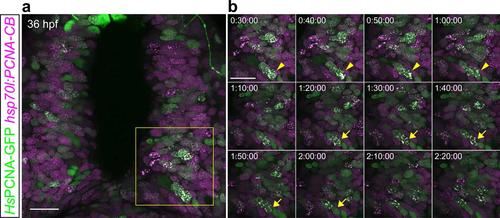
PCNA-CB and human PCNA-GFP dynamically colocalize when expressed in live zebrafish. 36 hpf hsp70l:PCNA-CB embryos transiently expressing human PCNA-GFP from an injected DNA construct. (a) Overview of the scanned area. (b) Detailed time lapse analysis. Live imaging of animals heat-shocked at 24 hpf shows that PCNA-CB and human PCNA-GFP mark the same nuclear structures and their pattern is characteristic of S phase. Additionally, both fluorescent fusions can trace these structures’ dynamics (arrowheads and arrows indicate two different cells over time). Scale bars: 20 µm. Timestamps: h:min:s. |
Expression Data
Expression Detail
Antibody Labeling
Phenotype Data
Phenotype Detail
Acknowledgments
This image is the copyrighted work of the attributed author or publisher, and
ZFIN has permission only to display this image to its users.
Additional permissions should be obtained from the applicable author or publisher of the image.
Full text @ Development