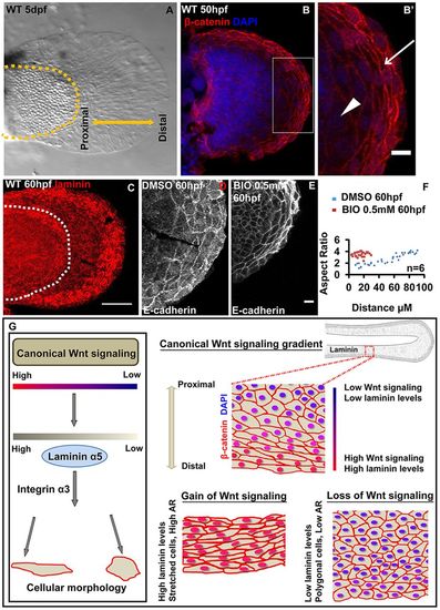
Mechanisms patterning median fin fold epithelium are also conserved in the pectoral fin. Bright-field image (A) of 5dpf wild-type pectoral fin. β-Catenin and DAPI-stained pectoral fin (B) and its digitally zoomed (B′) region (box in B). Arrow in B′ indicates stretched epithelial cells at the distal end, whereas arrowhead indicates polygonal cells. (C) Maximum intensity projections of wild-type larval pectoral fin stained for laminin. Confocal images showing E-cadherin staining in DMSO- (D) or BIO-treated (E) larval pectoral fins. (F) Comparison of aspect ratios of pectoral fin epithelial cells in DMSO and BIO-treated larvae. (G) Model showing the regulation of epithelial patterning in zebrafish fin-fold epithelium by the canonical Wnt signalling gradient. A gradient of canonical Wnt activity exists along the PD axis of the fin fold. The gain- and loss-of-function analyses reveal that the extent of canonical Wnt signalling regulates the expression of the ECM component laminin α5, which directly regulates cell shapes by interacting with integrin α3. Scale bars: 10 μm in B′,D,E; 50 μm in B,C. AR, aspect ratio.
|
