
Fig. 2
- ID
- ZDB-FIG-150629-11
- Publication
- Nagendran et al., 2015 - Canonical Wnt signalling regulates epithelial patterning by modulating levels of laminins in zebrafish appendages
- Other Figures
- All Figure Page
- Back to All Figure Page
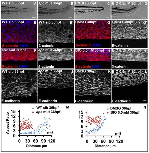
Gain of Wnt signalling results in acquisition of stretched cell morphology in the median fin fold. Bright-field images of wild-type sibling (A), apc mutant (B), DMSO- (C) and 0.5 mM BIO-treated (D) embryos. (E-H′) β-Catenin-DAPI overlays (E-H) and β-catenin staining (E′-H′) in given genetic backgrounds and treatments. (I-L) E-cadherin staining in wild-type siblings (I), apc mutant (J), DMSO control (K) and BIO-treated (L) embryos. (M,N) Comparison of epithelial cell aspect ratio plots between sibling and apc mutant (M) as well as between DMSO and BIO-treated embryos (N). Scale bars: 0.1mm in A-D; 10 μm in E-L. |
| Antibodies: | |
|---|---|
| Fish: | |
| Condition: | |
| Anatomical Terms: | |
| Stage Range: | Prim-15 to Prim-25 |
| Fish: | |
|---|---|
| Condition: | |
| Observed In: | |
| Stage Range: | Prim-15 to Prim-25 |