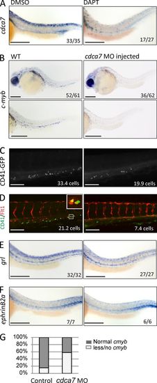
HSC formation is impaired in cdca7 morphants. (A) cdca7 ISH of representative embryos treated at 6 hpf for 24 h with DMSO or 100 μM DAPT. (B) Representative embryos (top) and detailed images of AGM region (bottom) from embryos treated with control or cdca7 MOs. Hematopoietic cells were detected by cmyb ISH at 30 hpf or by CD41-GFP at 50 hpf (C). The number of CD41-GFP–positive cells is significantly reduced upon cdca7 MO injection (P = 0.01). (D) Representative images from CD41-GFP and Flt1-RFP embryos treated with control or cdca7 MOs. Number of double-positive cells is indicated. A detail of CD41-GFP+/Flt1-RFP+ HSCs is depicted in the inset. (E and F) The dorsal aorta integrity is revealed by gridlock (hey2) and EphrinB2a expression in both WT and cdca7 MO-injected embryos. The numbers in the panels indicate the number of embryos of the total pool that displayed phenotype or the number of positive cells. (G) The percentages of cmyb expression are indicated in the graph. Bars: (A, B, E, and F) 200 μm; (C and D) 100 μm.
|
