
Fig. 1
- ID
- ZDB-FIG-150511-14
- Publication
- Bolcome et al., 2008 - Anthrax lethal toxin induces cell death-independent permeability in zebrafish vasculature
- Other Figures
- All Figure Page
- Back to All Figure Page
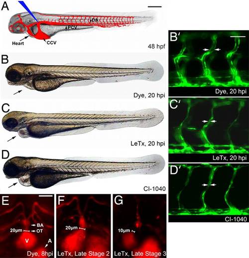
LeTx effects in zebrafish embryos. (A) Diagram of zebrafish embryo at 48 hpf (time point of toxin protein microinjection, at blue arrow), functional vasculature in red. (B–D) Images were taken at 68 hpf. (B) An inert phenol red dye was injected as vehicle in all control embryos and a WT phenotype was observed at 68 hpf, or 20 hpi. (C) 1× LeTx (defined below) generated pericardial edema and narrowed vessels (C′). (D) Embryo was treated with 2.5 µM CI-1040 at 48 hpf for 6 h, then washed out. Black arrow indicates heart and pericardial edema in A–D. (Scale bar, 250 µm.) (B2–D2) Enlarged images showing LeTx and CI-1040 induced narrowing of ISVs in Tg(fli1:EGFP)y (19) embryos, indicated by arrows. (Scale bar, 80 µm.) (E–G) LeTx-injected Tg(gata1:dsRED) (21) embryos exhibited outflow tract lumen size reduction from 20 µm to <10 µm between LeTx phenotype stages 2 (F) and 3 (G). (Scale bar, 50 µm.) DA, dorsal aorta; PCV, posterior cardinal vein; CCV, common cardinal vein; ISV, intersegmental vessel; BA, bulbous arteriosus; OT, outflow tract; A, atrium; V, ventricle, for all figures. 1× LeTx is defined as 37 fmol of LF and 25 fmol of PA. |